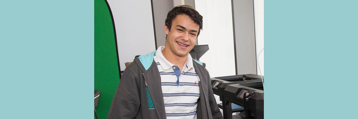
Nathan Bentolila - YULA Boys Class of 2016
This past year I have worked in two microscopy and imaging labs at UCLA. One was in the department of Physics and Astronomy and that other was at Californian Nano Systems Institute (C(N)SI) at UCLA.
Elegant Mind club Lab: Department of Physics and Astronomy, UCLA:We studied the behavior and anatomy of C. Elegans (a free-living (not parasitic), transparent nematode (roundworm), about 1 mm in length, that lives in temperate soil environments.). C. Elegans are very good animal model to better understand concepts and phenomenons that also occur in the human body. They have many of the same organs but on a much simpler and smaller scale, making it much easier to study.
During my time in the lab I worked on a few microscopes and projects. I learned how to navigate and program in both LabView and Matlab (Lab-view is used to control lab equipment and mat-lab is used to processes the data from the microscopes). Later on during the summer I designed and built my own microscope. My microscope's primary purpose was to be able to visualize the neurons (tagged with GFP) in the newly breaded worms. We did such to be able to ensure that the florescent gene was being passed on to the next generation of worms.
The Structure of My Microscope:
The microscope that I built is called a stereo-microscope or dissection microscope. This type of microscope uses a white light source which is then collimated through a series of lenses. The light beam is then passed through a blue light filter (which blocks all other colors except for blue). This blue light is then exposed to the sample which then, if present, stimulate the GFP in the worm causing it to glow. The light is then reflected off a mirror through an objective lens (20x or 10x) and through a green filter (only keeps the green rays). This allows one to visualize the green glow of the GFP. However in order for this to work I had to find a way to concentrate all of the light onto our sample. I accomplished this by using an illumination method called Köhler illumination. Köhler illumination uses a diaphragm and condenser lens to centralize the light onto the sample. Once I implemented this onto our microscope we were able to maximize the light passing through the filters and stimulating our sample. My microscope is now being used on a daily basis to visualize the GFP on the neurons of the C. Elegans. Using my microscope we were able to image two different strains of the C. Elegans worms (ST2 and QW1217). The ST2 has thousands of marked neurons (with GFP) making it fairly easy to image. However the QW1217 is usually a lot harder to image with a white light dissection microscope because the GFP is only tagged on 100 neurons. However after a lot of work we were able to image this strain as well. This is really amazing because before we were using much larger and more complex microscopes to image this strain.
Californian Nano Systems Institute (C(N)SI), UCLA:
I am currently working with a team of researchers who are studying melanoma in a mouse model. We are using the mouse’s ear to study melanoma because it is the easiest to image. The mouse’s ear is only a few cells thick which allows us to image the tumors. We are using an imaging method called Two-Photon Microscopy to image the tumors in the mouse’s ear. Two-photon excitation microscopy is a fluorescence imaging technique that allows imaging of living tissue up to about one millimeter in depth.
My primary role has been to design a specialized device to hold the mouse. I have developed and designed a product, which enables us to hold and stabilize the mouse for imaging (printed in the genesis lab). In past methods used, the mouse would move and the heartbeat of the mouse would distort the images. I am currently perfecting my design to suit the changing needs of the study. My product is in current use today in the lab’s active melonoma research.
